Home > Treatments > Treatments and Examinations > Conventional Examinations >
<
  • Color Doppler Ultrasound

    A A A
    Color Doppler Ultrasound
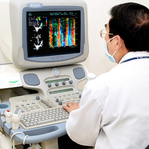
    As a kind of non-invasive medical imaging technology, it plays an important role in diagnosis and treatment of tumor. In terms of normal color doppler ultrasound, except early lung and brain tumor, it has a good effect on early detection and early treatment for tumors on the whole body. It is especially sensitive to tumors on thyroid, breast, liver and gall, pancreas, spleen, kidney, uterus, ovary and abdominal cavity. Color Doppler ultrasound is used in primary diagnosis on benign and malignant tumors and provides the clinicians the detailed information on relationship between tumor size, shape, location and peripheral tissue, and the scope of the tumor invasion, the diagnosis of metastasis and melanoma.

    The development of the multi-color Doppler ultrasounds new technology plays a great role on tumor detection and treatment. For example, technologies such as contrast-enhanced ultrasound, elastography, ultrasound-guided puncture and biopsy is more accurate to detect whether a tumor is malignant or not. Ultrasound-guided tumor puncturing interventional therapy such as freezing, radiofrequency, microwaves and radioactive particles play a very important role in treatment of the focal. Aside from cancer detection and diagnosis, color doppler ultrasound is also helpful to cancer patients in other aspects. For example, it is not only used in check up and monitoring on cancer patient’s heart function, but also plays a very important role in helping clinicians make more appropriate treatments planning for cancer patients.
    
    
  • Capsule Endoscopy

    A A A
    Capsule Endoscopy
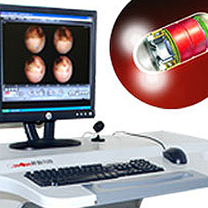
    Capsule Endoscopy is a new era in the history of endoscopic technique. Capsule Endoscopy technique develops very fast, which begins from diagnosis of obscure gastrointestinal bleeding, to screening intestinal diseases. It can also make correct diagnosis and differential diagnosis for obscure abdominal pain, diarrhea, hemorrhage, anemia, and weight loss.

    The whole capsule endoscopy system consists of three parts, including capsule/signal transmission, signal receiver/recorder and image workstation. Capsule Endoscopy can be used in gastrointestinal screening, but mainly in small intestine inspections. It can also be used in inspection of inflammatory bowel disease, parasite, vascular, congenital heteroplasia, and drug and radiation injury, especially small intestinal tumor. Small intestinal tumor accounts for 5% in gastrointestinal tumor. On the one hand, the incidence rate of this disease is comparatively low. On the other hand, it is concerned with the immaturity of the medical diagnosis method. Small intestinal diseases are detected mainly by Enteroclysis, but its positive rate of diagnosis is very low. CT scanning can also detect small intestinal tumor, and can detect primary tumor and metastasis, but the detection rate for small lesion and mucosal lesion in hollow organ is very limited.

    With the development of Endoscopic technique, Endoscopic screening (Enteroscopy and capsule Endoscopy) has developed into the best diagnostic tool. There are some limitations on Enteroscopy, which belongs to invasive detection. The patient needs anesthesia before receiving detection and it also has disadvantages like expensive operating fee, low popularity, and poor patient compliance. With the application and emergence of capsule Endoscopy, the detection rates for small intestinal tumors have increased obviously. Compared with Enteroscopy, capsule Endoscopy has the advantages such as non-invasive, lower cost and the patient do not need anesthesia. Small intestinal tumors include small intestinal benign tumor and malignant tumor. The former one includes various kinds of polyps, adenoma, leiomyoma, lipoma, hemangiomas, hamartoma, lymphangiomyoma, neurofibromas, etc. The latter one includes gastrointestinal stromal tumors, adencarcinoma, neuroendocrine carcinoma (carcinoid and small cell neuroendocrine carcinoma), lymphoma etc. Capsule endoscopy provides significant basis on diagnosis and treatment for small intestinal tumors. Many malignant tumors such as melanoma, breast cancer, lung cancer, renal cell carcinoma, osteosarcoma, and germ cells cancer can also transfer to the small intestine. It should be paid more attention in clinical work. Capsule Endoscopy has a very wide range of applications in clinical work.
    
    
  • Gastroscope

    A A A
    Gastroscope
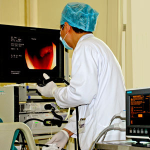
    Since 1868, metal pipe gastroscope has been applied to examine the patient. With more than one hundred years evolution and development, gastro-fiberscope has emerged. In particular, electronic gastroscope emerged in 1982, which inaugurated a new era in gastroscope history. Electronic gastroscope has a number of advantages such as a soft lens body, easy manipulation, less pain, low risk, clear image. Gastroscope has a very wide range of indications including suspected upper gastrointestinal tract lesions, abdominal discomfort; ulcer, tumor and other lesions through X-ray examination, acute gastric hemorrhage, chronic and unknown blood loss; follow-up on different kinds of upper gastrointestinal tract lesions such as ulcer, atrophic gastritis, postoperative stomach; gastroscope can make correct diagnosis especially for stomach neoplasms (benign or malignant) lesion location, size, shape, and depth; also can dynamically observe the change of lesions and provide the basis for surgery.

    Gastroscope can be used for some special examinations such as biopsy, cytological examination, bacteriology examination, mucosa staining examination, functional test & then photography operations and lesions. A biopsy under direct vision is one prominent advantage of gastroscope. During this process, if putative lesions are identified, conduct tissue biopsy and pathological examination to confirm diagnosis. Smear and cytological examination has significant effect for diagnosis in malignant tumors. For those with severe luminal stenosis, as the gastroscope cannot reach the lesion, cytologic smear is very important, making up for a lack of biopsy and it is particularly important for those with severe luminal stenosis, as the colonoscopy cannot reach the lesion. In the aspect of bacteriology examination, the relationship between helicobacter pylori (Hp) and gastropathy draws the society's attention. WHO has defined helicobacter pylori (Hp) as group one carcinogen. Hp inspection can provide some reference for diagnosis and treatment of gastropathy. The adopters for mucosa staining method presently are C.I.Acid blue 74, methylene blue, congo red, tattoo and Iodine solution. Functional examination makes the endoscopic diagnosis get rid of observing the shape and histopathology solely with the naked eye. It can observe stomach motion, the blood flow and the changes of pH level. At present, gastroscopy can be done with no pain, which makes the patient complete the examination in a comfortable state.

    Gastroscopy has achieved fast growth, widening more treatment field. In the aspect of endoscopic therapy, it also makes great progress at home. Endoscopic therapy can solve problems which involved surgery before.

    High frequency technology: High frequency current can produce high temperature, promoting cell differentiation and proteolysis, which has effect on the opening, freezing, and occurrence of spark, widely used in treatment of stomach polyps.

    Microwave Therapy: It is only used in thermotherapy from the beginning and then used in freezing, cutting and hemostasis of the tissue. It is also used in treatment of early gastric cancer and removal of cancer stenosis and stomach polyps.

    Laser Therapy: Similarly, it can cause tissue gastification and freezing and is applied in cutting and hemostasis.

    Drug Injection: It can be applied in treatment of esophageal cancer through anti-cancer drug injection, to remove of cancer stenosis and can partly relieve the patient’s symptoms.

    Endoscopic Mucosal Resection: It is a new technology widely applied in clinics. It is applied in diagnosis of the lesion that is difficult to find through routine biopsy, and applied for removal of early gastric cancer and precancerous lesion.
    
    
  • Colonoscopy

    A A A
    Colonoscopy
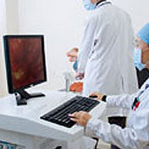
    After 50 years of development, electronic colonoscopy has been widely applied in clinics. Colonoscopy is indicated to hematochezia of unknown origin, diarrhea, non smooth defecation, changes in bowel habits, constipation, stomachache and abdominal mass. It can make diagnosis of all kinds of large intestine disorder and it can precisely determine on the property (benign or malignant), size, shape and stage of the tumor. Moreover, colonoscopy provides clues for treatment, especially for surgery, as it can observe the lesion changes dynamically.

    Colonoscopy is also available for biopsy, cytological examination, bacteriology examination and mucosal staining, and the operation process and the condition of lesion will be recorded. Conducting biopsy under direct vision is an outstanding advantage of colonoscopy. If suspicious lesions are found, conduct tissue biopsy and pathological examination to confirm diagnosis. Cytologic smear is vital to diagnosing of malignant tumor and it can cover the shortage of biopsy. For those with severe  luminal stenosis, as the colonoscopy cannot reach the lesion, cytologic smear is  particularly important. The colonoscopy can be done with no pain.

    In the treatment guided by gastrointestinal endoscopy, the high frequency electric energy is commonly used to remove the intestinal polyps. The high frequency electric energy, microwave and laser can be applied to treat various types of bleeding and relieve neoplastic stenosis. The endoscopic mucosal resection is a new technique  with extensive development in recent years. It is indicated to the biopsy of undiagnosed lesion in conventional biopsy, resection of early-stage intestinal cancer and large, benign and ridgy lesion
    
    
  • CT

    A A A
    CT
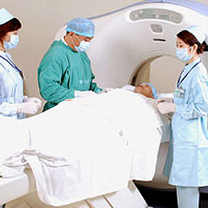
    CT scan is of significant value in diagnosing diseases on the whole body and is the important basis for diagnosing cancer diseases. CT exam is of high value in diagnosing central nervous system diseases, which can be applied widely in intracranial tumor, injury and infection etc. CT scans can specifically identify brain abscess, parasitic infections, traumatic hematoma, brain injury, cerebral infarction, cerebral hemorrhage and brain tumors etc. It also has good diagnostic effect on intra-spinal tumour and lumbar intervertebral disc extrusion. Furthermore, CT scans are of high value of otolaryngologic and neck diseases such as orbital lesions, paranasal sinus cancer, cholesteatoma of middle ear, parotid tumor, nasopharyngeal cancer etc.

    CT scan is of great value in diagnosing chest diseases, especially for spiral CT. The diagnosis for chest diseases has shown its advantage day by day. Using enhancement technology can specify whether lymphadenopathy and lung tumor or lymphoid tissues enlarge abnormally. It can also help diagnose lung lesions, bronchus narrow or obstruction. CT is helpful in differential diagnosis of primary or metastatic tumors, scrofula, central lung cancer, etc.

    64-slice spiral CT is of great value in diagnosis of heart and cardiovascular diseases, which can clearly diagnose if the patient has coronary artery calcification or myocardial bridge. Coronary artery CTA can clearly diagnose if the patient has coronary artery stenosis and the narrow degree which can also diagnose artery diseases such as aortic dissection, aortic aneurysms, aortic intramural hematoma, ulcer etc. It provides great value for early diagnosis and treatment of cardiovascular diseases.

    CT scan is widely used in abdomen and pelvis, which is used mainly in diagnosis of the liver, gallbladder, pancreas, spleen, retroperitoneal space diseases. It is of great value for urogenital diseases and can clearly diagnose space-occupying lesion, inflammation, and injury etc. CT can help to diagnose gastrointestinal cancer and detect if the tumor compress adjacent organs and distant metastasis of the tumor.

    Notice: CT scan is generally not recommended for women in early pregnancy.
    
    
  • MRI

    A A A
    MRI
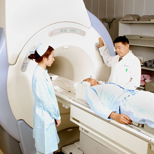
    MRI is of great value in diagnosis of head, spine, and spinal cord diseases. The diagnosis for joint parenchyma trauma such as meniscus, cruciate ligament injury is better than CT scans. It also has great value in diagnosis of otolaryngologic disease, heart disease, mediastinal mass, osteoarthrosis, muscle disease, abdominal organ (such as liver, spleen, pancreas, kidney, adrenal gland) and pelvic organ(such as uterus, ovary, bladder, prostate) lesion. MRI is also of significant value in early diagnosis in prostate cancer and has no radiation injury on the human body.
    
    
  • PET/CT

    A A A
    PET/CT
    PET/CT is the ideal combination between nuclear medicine equipment and single photon emission computed tomography in the recent years. With the rapid development of computer science and biology, PET/CT is applying widely on the study of oncology.

    1.Tumor screening: According to statistics, in People’s Liberation Army General Hospital, subclinical cases of malignant cancer reach 2% among one thousand asymptomatic population who get heath exam and their CT and MR exam shows normal findings.

    2.Qualitative diagnosis of tumors: Tumor cell has high uptake rate for (18)F-FDG, which can reflect division and proliferation of malignant tumors or neovascularization of tumors.

    3.Cancer staging: Staging is a way of describing where a cancer is located, if/or where it has spread, and whether it is affecting the functions of other organs in the body. Doctors often use diagnostic tests to determine a cancer’s stage, so staging may not be complete until all of the tests are finished. It will help to achieve tumor individual therapy.

    4.Tumor biological characteristics. The concentration degree of tumor cell for tracer directly reflect tumor malignancy.

    5.Unknown primary tumor testing: It is very common to first detect metastatic tumors with unknown primary site in clinic. PET/CT can discover the primary tumor through a whole body scan and also provide the scope and biological characteristics of the tumor.

    6.Assistance for tumor treatment planning. During the process of tumor radiotherapy, PET/CT can hint the distribution of bioactive tumors to facilitate making of the radiation treatment plan.

    7.Monitoring and prediction of tumor treatment: PET/CT is used in early detection of the sensitivity of tumor cell for radiotherapy or a certain kind of chemo-therapeutant, which can facilitate the modification of individualized therapeutic scheme according to treatment effect.

    8. Monitoring & Restaging of tumor recurrence

    9. New technology & drug research on tumor treatment
    
    
  • SPECT

    A A A
    SPECT
    SPECT is the nuclear medical equipment and single photon emission computed tomography. It is an important tumor diagnosis equipment before PET/CT is developed. It is still an irreplaceable equipment in the diagnosis of the whole body bone metastasis of different malignant cancer. By the first imaging method, it can detect bone metastasis that other equipments can not detect and plays an important role in making individualized treatment plan. SPET also has great value in dynamic checking of myocardial ischemia of coronary heart diseases & kidney function.
    
    
  • Blood Examination

    A A A
    Blood Examination
    Blood examination is one of the important means to detect early cancer, which can detect various tumor marker indexes, find and detect various malignant cancers. For example, when carcinoembryonic antigen(CEA) rise obviously, the patient may suffer from colon cancer, gastric cancer, lung cancer and cholangio carcinoma. If liver cancer, breast cancer, ovarian cancer, pancreatic cancer exists, it may also rise.

    Tumor markers are substances found at higher than normal levels in the blood of some people with cancer. A sample of blood will be taken and then sent to a laboratory for analysis.
    
    
  • Mammograph

    A A A
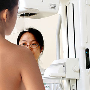
    Mammograph
    Breast Mammograph, its full name is breast mammograph X-ray photography exam, also called Mammograph. It is the first choice in the diagnosis of breast diseases and the simplest, reliable and noninvasive inspection method. Its characteristics are to clarify the chest mass that the doctor can not touch. The agony of the patient is comparatively small and easy to cooperate. With high resolution, good repeatability, the stored pictures can be used for comparison and are unrestricted to the age and body shape. At present, it has been a kind of routine exam.

    Breast mammograph has become one of best ways for clinical routine exams and preventive inspection for breast cancer because it features comprehensiveness, intuitive, easy-to-use, safety and requires a low cost. It makes contribution to detecting early carcinosis, improving breast diseases diagnosis coincidence rate, and survival rate of patients.
    
    
>

Conventional Examinations


Treatment Overview
Treatments
Technological Advantage
Treatment Strategy

TOP


Clifford Hospital is the first Chinese hospital accredited by Joint Commission International (JCI), and has passed 5 times of JCI accreditation.

Copyright © 2013-2015 by Clifford Hospital-A JCI Accredited Hospital, Level A Tertiary Hospital, All Rights Reserved.
Technical support:Guangzhou Fortune Software Co.,Ltd  Sitemap