Home > Treatments > Treatments and Examinations > Other Treatments >
<
  • Cryoablation Therapy

    A A A
    Cryoablation Therapy
    The cryocare TM targeted cryoablation therapy is also called argon-helium knife. It employs the space rocket guidance technology and the oncology theory of modern ultra-low temperature cryotherapy. The refrigerant (argon) can freeze the lesion to -140℃ while the thermal medium (helium) can heat it up to 45℃. This process takes no more than 50s and the combination of argon and helium destroys and kills cancer cells. Because of the destruction of cancer cell, the antigen of cancer cell enters into the blood and boosts the immune system to generate the cellular and humoral immune response, thus improving the immunity.

    As the argon-helium ultra-cold knife is a minimally invasive treatment and it can  completely eradicate the tumors, it can be applied to the patients including post-surgery recurrent patients, elder people, people in weak condition, people unwilling to undergo surgery, patient with metastasis, cancer patient with aortic surrounding tissues that cannot be removed through surgery, patients with poor therapeutic effect of radiotherapy, chemotherapy and other therapies or patient no longer sensitive to those therapies, patients that suffer from treatment failure. The cryoablation therapy can minimize the tumor burden, quickly relieve pain, greatly improve the therapeutic effect of immune therapy, traditional Chinese medicine, radiotherapy and follow-up care, reduce side effect, effectively extend life span and improve the quality of life.


  • Gamma Knife Surgery

    A A A
    Gamma Knife Surgery
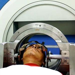
    Gamma Knife is also known as stereotactic gamma-ray radiation therapy. It employs modern computer techniques, stereotactic technique and surgical technique. Gamma Knife is not a normal scalpel. It uses beams of geometrically-focused gamma rays to focus on the target through precise stereotactic position and a certain dose of gamma rays is delivered to the site where all the beams meet to massively destroy the targeted tissue at once. This result is similar to an actual surgical procedure. As the healthy tissues are in the outside of lesion and focused-on single beam of gamma rays, there will be no harm to healthy tissues in such low energy. By using a magnifying glass, the sunshine is narrowed to a highly localized area. This results in a concentration of heat that can reach incredibly high temperatures. If a high enough temperature is reached, the kindling will smolder and begin to burn. However, there will be no burning in the other area. This phenomenon can further explain how the gamma knife radiosurgery works. As there is distinct boundary between the range of radiation and healthy tissues, which seemed like cutting by a knife, this therapy is called gamma knife. Gamma knife has the advantages of non-invasiveness, no need for general anesthesia and actual surgery, no bleeding and inflammation.  

    Gamma knife can be divided into head gamma knife and body gamma knife. Head gamma knife is used to treat small tumors and other functional diseases in the brain while body gamma knife is mainly applied in treating all kinds of whole body tumors.

    Indications of Head Gamma Knife
    1.Brain tumors whose size is within 30mm, such as acoustic neuroma, pituitary adenoma, meningioma, pineal region tumor and lymphoma, etc.

    2.Cerebral arteriovenous malformation (AVM)

    3. Cavernous hemangioma

    4.Benign tumors that cannot be completely eradicated through surgery

    5. Intracranial metastatic carcinoma of small size and clear tumor demarcation

    6. Functional neurological disorder like primary prosopalgia or prosopalgia that fail to be treated with drug, intractable pain, dyskinesia, epilepsy and other functional diseases caused by Parkinson's disease.

    7.Spinal cord tumor of cervical spinal nerve 7 (C7) and above

    8.Extracranial head and neck tumors

    9.Senior citizens, patients with chronic diseases like diabetes, hypertension, heart disease, people that are weak, brain cancer patient that can’t tolerate surgery and conventional radiotherapy.

    Indications of Body Gamma Knife
    Indications of body gamma knife include nasopharyngeal cancer, laryngeal cancer, thyroid cancer, primary and metastatic lung cancer, primary and metastatic liver cancer, pancreatic cancer, gallbladder cancer, bile duct cancer, metastatic cancer of lymph node in neck, hilum of lung, mediastinum, abdominal cavity and pelvic cavity, esophagus cancer, gastric cancer, mediastinal tumor, kidney cancer, rectal cancer, bladder cancer, prostate cancer, lymphoma, malignant thymoma, gynecological tumors like cervical cancer and ovarian cancer, primary bone tumor, bone metastasis and all kinds of sarcomas.

    Gamma knife radiosurgery can be a locally radiotherapy boost treatment for the residual tumors and serve as an alternative radiotherapy for treating recurrence after the patient finish taking the conventional radiotherapy. 
    
    
  • Radio Frequency Ablation Therapy

    A A A
    Radio Frequency Ablation Therapy
    Concept
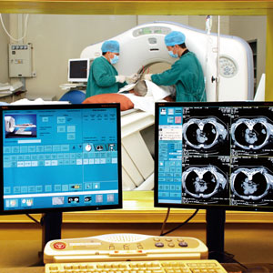
    There is growing concern for radio frequency ablation (RFA) as it is the “new shining star” in minimally invasive cancer treatment. Conventional radio frequency ablation finds the tumor through ultrasound, CT and MRI. Guided by imaging techniques, the doctor inserts a thin needle (electrode) with the length of 15 to 20 cm through the skin and into the tumor. High-frequency electrical energy delivered through this needle heats and destroys the tumor, thus causing coagulative necrosis of cancer cell. Radio frequency ablation can be effective for liver tumor, lung tumor and kidney tumor, etc.

    Therapeutic Effect
    1.Kill cancer cells rapidly: The radiofrequency waves released by needle electrode make the polar molecules in the tumor tissues to undergo rapid changes in the direction of the radiofrequency alternating current. The produced frictional heat causes the temperature of treating area to rise to 90 to100℃, even more than 100℃ in a matter of minutes. As the heat tolerance of cancer cells is much lower than that of normal cells, cancer cells can be effectively and rapidly killed in 42℃ or above, then heat cogulative necrosis occurs.

    2.Prevent tumor metastasis: Radio frequency ablation can clot the blood vessels of tumor tissue, which is good for tumor metastasis prevention.

    Indications
    1.Primary lung cancer, metastatic lung cancer, pleural tumor, metastasis in the bone of chest wall and sole tissue, mediastinal tumor and etc…

    2.Patients who can’t tolerate surgery, unwilling to undergo surgery or with high risk of surgery and senior patients.

    3.Treatment for recurrent cancers and metastatic liver cancer that is unavailable to undergo surgery.

    4.Remedy for surgical exploration: lung tumor with chest wall invasion; metastatic tumors in the chest walls.

    5.The distance between lesion and great vessels and trachea is over 1 cm.

    Advantages
    1.Strong safety. With the guidance of advanced equipment, this therapy can keep away from the nerve and vessels and precisely target the lesion without injuring the healthy tissue and peripheral nerve.

    2.Remarkable therapeutic effect. For the tumor diameter less than 5cm, especially the liver tumor in depth, taking the radio frequency ablation once can eliminate it. This therapeutic effect is as good as that of surgery. For the tumor diameter more than 5cm, this therapy can reduce the risk of surgery.

    3.Minimally invasive. No need for surgery, little injury, quick recovery, short inpatient time and low costs.

    4.Few complication. Unlike surgery, there is no danger for introgenic residual and intrahepatic metastasis in radio frequency ablation and the radio frequency ablation can clot the blood vessels of the tumor tissues, this in turn is good for tumor metastasis prevention.

    5.Wide range of application. It can be applied to major primary, recurrent and metastatic cancer.   


  • Particle Implantation Therapy

    A A A
    Particle Implantation Therapy
    Concept
    Particle implantation therapy is performed under the guidance of medical devices. In this therapy, doctors implant miniature radioactive particle in the tumor and the  particle is proven to have the effect of killing cancer cells. Then the persistent and short-wave radiation released by miniature radioactive source kills the cancer cell and allows the tumor to shrink.

    Particle implantation technique falls in the category of minimally invasive brachytherapy, which can effectively kill the cancer cells and protect the surrounding healthy tissues from damage. As for the advanced cancer patients, particle implantation can prolong their survival time, improve quality of life and control cancer pain. As for the early-stage and medium cancer patients, it can greatly improve the treatment process.

    Advantages
    1.The particle implantation therapy is well-targeted with remarkable therapeutic effect. The radioactive particle implanted in the tumor can intensively and effectively kill cancer cells and allow the tumor to shrink or even disappear. 

    2.Minimal damage and no complication. The particle implantation damages the cancer cell within the 1-cm distance of the radioactive source and does not harm other tissues, which can effectively improve the cancer patient’s quality of life.

    3.Small wound. The radioactive particle is usually implanted under the guidance of B-ultrasound and CT, or during surgery. There is a small wound and a relatively light pain.

    4.Effective and comprehensive treatment. The particle implantation therapy can integrate with other therapies like surgery and chemotherapy to improve the cure rate of surgery and reduce the course of chemotherapy.

    Indications and Contraindications
    Indications
    1. Untreated primary tumor such as prostate cancer and so on.

    2. Important functional tissues that need to preserve important organs that may be involved in surgery, like the tumors deep within the brain.

    3.Cases of unwillingness to undergo radical surgery, such as thyroid cancer, endometrial cancer, and cervical cancer and so on.

    4. Metastatic lesion or postoperative solitary metastatic lesions that are not candidates for surgery, like the multiple metastatic lung tumor.

    5. Unresectable primary cancer, such as large mass liver cancer, nasopharyngeal cancer and so on.

    6.New version: Radiotherapy boosts treatment for the locally residual tumors. Iodine-125 implantation can be performed.

    7.Poor therapeutic effect or failure case of radiation
     
    Contraindications
    Bleeding disorder, organ failure and mental disorder.
    
    
  • Photodynamic Therapy

    A A A
    Photodynamic Therapy
    Photodynamic therapy (PDT) is a new technology that uses photodynamic effect in diagnosis and treatment. This therapy is based on the photodynamic effect. The photodynamic effect is a photosensitized reaction accompanied by the biological effects and getting the molecular oxygen involved. In the process of photodynamic effect, laser radiation at specific wave length makes the photosensitizer absorbed by the tissue become excited, and then the transfer of energy from the excited photosensitizer produces the highly reactive singlet oxygen. The singlet oxygen can react with neighboring biomacromolecule to generate oxidation reaction, which results in cytotoxic effects, leading to the damage and death of the cell. Many hospitals have applied the photodynamic therapy in the diagnosis and treatment for cancer so far. Additionally, many organizations and institutes are studying this therapy.

    The Indications of Photodynamic Therapy:
    It is applied to the orificial, cavitary, body-surface tumors and abnormal proliferation

    Major indications: esophageal cancer, gastric cardia cancer, bronchogenic carcinoma,  bladder cancer and nasopharyngeal cancer and etc…

    Other indications: gastric cancer, oral cancer, tongue cancer, laryngeal cancer, bronchial cancer, lung cancer, bile duct cancer, liver cancer, cervical cancer, colorectal cancer, skin cancer, breast cancer, brain cancer, vaginal cancer, penile cancer, encephalic glioma, and etc…

    The photodynamic therapy can also indicate to the following types of skin cancer: basal-cell carcinoma, squamous cell carcinoma, malignant melanoma, Bowen's disease, Paget's disease and carcinoma in situ and etc...

    This therapy is especially suitable for the senior patients, those with contraindications to surgery, chemotherapy and radiotherapy, and patients with relapse after surgery or radiotherapy. 
    
    
  • German WOLF Laparoscopy

    A A A
    German WOLF Laparoscopy
    Concept
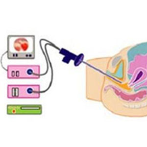
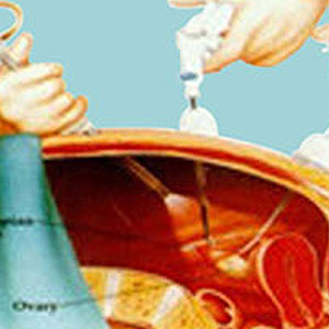
    The laparoscopy is one of the minimally invasive surgical techniques. During laparoscopy, the surgeon makes one or more “key hole” an incision around surgical site through which laparoscope is placed so the surgeon does not need to perform laparotomy. Moreover, the surgeon can look directly at contents of a patient's abdomen or pelvis on a computer screen and perform accurate surgery. The process only lasts for a short time. This laparoscopy represents the epoch-making progress of conventional laparotomy. 

    The Advantages of Laparoscopy
    Utilizing the laparoscopy inspection technique, the doctor can conduct inspection and treatment on patient’s womb and abdominal cavity at the same time. For example, when the doctor inspects something abnormal like endometrial polyp and fibroid, he can operate surgery for the patient, which saves both time and money. The specific advantages are as follow:

    1. Accurate determination: The laparoscopy is a high-tech minimally invasive medical instrument and it can achieve the goal of accurate determination in the lesion within the womb.

    2. No open procedures: Compared with the conventional laparotomy, the laparoscopy procedure requires no open procedures. Also, there are no wound and pain as well.

    3. Uterus preservation: As for patients with tumors the diameter of more than 6cm, the laparoscopy can preserve their physiological function and patients’ mental state will not be affected whatsoever. 

    4. Quicker recovery times: Patient’s hospital stay lasts for 2 to 3 days. There is a few complications in the short term while no ovarian function affected in the long term.

    5. Slight damage: Small incisions, whose size is 0.5cm; slight damage during the operation; short operation time, less bleeding and there will be no worm-like scar on the abdomen.

    6.Less pain: Only 3 small incisions are made on the belly and abdomen; no incision of diseased organ so the healthy tissue won’t get harmed and there is less pain after surgery.

    Indications
    Radical cure for early gynecological cancer, such as cervical cancer, ovarian cancer, endometrial cancer, pelvic cancer, fibroid, ovarian cysts, adnexal cysts, chocolate cysts, vaginal cancer, uterine cancer, uterine sarcoma, fallopian tube tumors, cervical intraepithelial neoplasia (CIN), ovarian teratoma, hysteromyoma and so on.

    As for the advanced gynecological cancer that loses the chance for surgery, the laparoscopy can solve many complications. A satisfactory therapeutic effect will be achieved when combining the laparoscopy with comprehensive techniques, such as traditional Chinese medicine, immune therapy and so on. 
    
    
  • High-Intensity Focused Ultrasound

    A A A
    High-Intensity Focused Ultrasound
    Concept
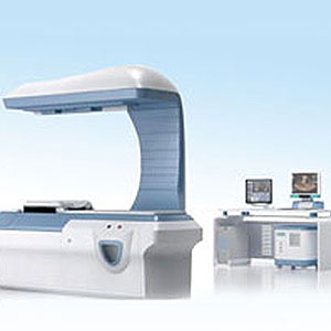
    High-intensity focused ultrasound, also called HIFU knife, is a new technique to kill tumors without surgery or puncture. It is regarded as an excellent, non-invasive localized physical therapy in vitro by domestic and international experts. Many large-scale hospitals both at home and abroad have promoted the use of high-intensity focused ultrasound. Tens of thousands of patients have taken this therapy, which received remarkable therapeutic effect.

    The HIFU treatment has many advantages: non-invasiveness, no requirement for anesthesia and puncture, no blood loss, remarkable therapeutic effect, quick recovery, strong security and repeated administration. Moreover, there is no restriction on the size and shape of the tumor. This treatment is an ideal way for people to perform surgery in vitro.

    Principle of Treatment
    HIFU knife treatment utilizes the ultrasound wave as a therapeutic source and it focalizes the power of ultrasound wave to destroy cancer cell with pinpoint accuracy, which causes coagulative necrosis of tumor tissue and makes it lose its ability to proliferate, invade and spread. This principle is similar to that of solar cooker, with concentrated sunlight generating great energy at the focus area. Generally speaking, the tumor will stop growing and be absorbed by the body gradually after the HIFU treatment.

    Advantages
    High-intensity focused ultrasound (HIFU knife) is regarded as an excellent, non-invasive localized physical therapy in vitro by oncological professionals. Its advantages are as follow:

    1.Effective treatment: Ultrasound wave can localize the cancer lesion in real time, and monitor the treatment process so it can effectively damage the cancer cell and ensure the therapeutic effect.

    2.Strong security: As a physical therapy, there is no surgical incision, ionizing radiation and radiation pollution in radiofrequency ablation. Patients do not need to undergo general anesthesia, so this treatment is safe and free of potential risks.

    3. Preservation on the organs and their function: As the radiofrequency ablation can damage the tumor tissue accurately and protect the healthy tissues, it is beneficial for preserving patient’s healthy tissue and its function of the cancerous organ. So it is possible for breast cancer patients to conserve their breast and for patients with malignant tumor of limbs to preserve their limbs.

    4. Slight damage and few side effects: The radiofrequency ablation is a non-invasive therapy in vitro. There is no infection during the treatment process. Patient has quick recovery time with no scar present after the treatment. Moreover, as the treatment is conducted in vitro, it avoids the iatrogenic implantation of cancer cells, which is good for localized control of the tumor and reducing the chance of metastasis.

    5. Strengthen patient’s immune function to fight cancer: After the high-intensity focused ultrasound treatment, the tumor tissue is damaged. Then the dead cancer cells release the tumor antigen and boost patient’s immune function to fight cancer .

    6. Strong effect of comprehensive treatment: The high-intensity focused ultrasound damaged primary tumor tissues and residual lesion from three aspects, which are killing the cancer cells with extreme heat, damaging the blood vessels that nourish the tumor, and strengthening the immunity to fight cancer after treatment. This treatment effectively treats tumor both in local and the whole body and achieves ideal effects of comprehensive treatment.

    7. Available to repeated administration: Local recurrence of tumor after surgery is one of the major causes of cancer treatment failure. For example, during the first surgery of large liver cancer, there may be removal of normal liver tissue in large numbers. So it will be difficult to conduct another surgery when there is local recurrence. However, when there is active cancer cell and cancer lesion in local verified by MRI after taking the HIFU knife treatment, patients are available to repeat administration till satisfactory effect has been achieved.

    Indications
    The high-intensity focused ultrasound is indicated to; extensive, primary and metastatic liver cancer, breast cancer, malignant bone tumors, kidney cancer, pancreatic cancer, soft tissue sarcoma, retroperitoneal tumor, pelvic tumors, bladder cancer, palliative treatment for advanced malignant tumor or metastatic tumor, fibroid, mammary fibroma, benign liver tumor and all kinds of solid tumors verified by the B-ultrasound. 


>

Other Treatments


Treatment Overview
Treatments
Technological Advantage
Treatment Strategy

TOP


Clifford Hospital is the first Chinese hospital accredited by Joint Commission International (JCI), and has passed 5 times of JCI accreditation.

Copyright © 2013-2015 by Clifford Hospital-A JCI Accredited Hospital, Level A Tertiary Hospital, All Rights Reserved.
Technical support:Guangzhou Fortune Software Co.,Ltd  Sitemap