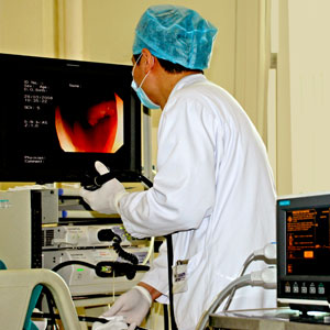
Since 1868, metal pipe gastroscope has been applied to examine the patient. With more than one hundred years evolution and development, gastro-fiberscope has emerged. In particular, electronic gastroscope emerged in 1982, which inaugurated a new era in gastroscope history. Electronic gastroscope has a number of advantages such as a soft lens body, easy manipulation, less pain, low risk, clear image. Gastroscope has a very wide range of indications including suspected upper gastrointestinal tract lesions, abdominal discomfort; ulcer, tumor and other lesions through X-ray examination, acute gastric hemorrhage, chronic and unknown blood loss; follow-up on different kinds of upper gastrointestinal tract lesions such as ulcer, atrophic gastritis, postoperative stomach; gastroscope can make correct diagnosis especially for stomach neoplasms (benign or malignant) lesion location, size, shape, and depth; also can dynamically observe the change of lesions and provide the basis for surgery.
Gastroscope can be used for some special examinations such as biopsy, cytological examination, bacteriology examination, mucosa staining examination, functional test & then photography operations and lesions. A biopsy under direct vision is one prominent advantage of gastroscope. During this process, if putative lesions are identified, conduct tissue biopsy and pathological examination to confirm diagnosis. Smear and cytological examination has significant effect for diagnosis in malignant tumors. For those with severe luminal stenosis, as the gastroscope cannot reach the lesion, cytologic smear is very important, making up for a lack of biopsy and it is particularly important for those with severe luminal stenosis, as the colonoscopy cannot reach the lesion. In the aspect of bacteriology examination, the relationship between helicobacter pylori (Hp) and gastropathy draws the society's attention. WHO has defined helicobacter pylori (Hp) as group one carcinogen. Hp inspection can provide some reference for diagnosis and treatment of gastropathy. The adopters for mucosa staining method presently are C.I.Acid blue 74, methylene blue, congo red, tattoo and Iodine solution. Functional examination makes the endoscopic diagnosis get rid of observing the shape and histopathology solely with the naked eye. It can observe stomach motion, the blood flow and the changes of pH level. At present, gastroscopy can be done with no pain, which makes the patient complete the examination in a comfortable state.
Gastroscopy has achieved fast growth, widening more treatment field. In the aspect of endoscopic therapy, it also makes great progress at home. Endoscopic therapy can solve problems which involved surgery before.
High frequency technology: High frequency current can produce high temperature, promoting cell differentiation and proteolysis, which has effect on the opening, freezing, and occurrence of spark, widely used in treatment of stomach polyps.
Microwave Therapy: It is only used in thermotherapy from the beginning and then used in freezing, cutting and hemostasis of the tissue. It is also used in treatment of early gastric cancer and removal of cancer stenosis and stomach polyps.
Laser Therapy: Similarly, it can cause tissue gastification and freezing and is applied in cutting and hemostasis.
Drug Injection: It can be applied in treatment of esophageal cancer through anti-cancer drug injection, to remove of cancer stenosis and can partly relieve the patient’s symptoms.
Endoscopic Mucosal Resection: It is a new technology widely applied in clinics. It is applied in diagnosis of the lesion that is difficult to find through routine biopsy, and applied for removal of early gastric cancer and precancerous lesion.


