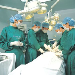
Oncological Surgeries of General Surgery Department
Except for conventional laparotomy, the following are therapies in cancer operation of General Surgery Department:
1.Minimally invasive operation
Endoscopic Operation: resection of gastric cancer, resection of colorectal cancer, resection of liver cancer, resection of pancreatic cancer, resection of thyroid carcinoma, etc.
Mammotome minimally invasive therapy of breast cancer.
2.Ablation
Physical ablation: It includes thermal ablation (microwave ablation and cryoablation).
Chemical ablation: This mainly refers to use/effects of drugs and other chemicals, such as ethanol injection.
3.Endoscopy operation
Gastroscope operation: Small benign tumor of the esophagus, gastric mucosa and canceration of the esophagus, gastric mucosa (carcinoma in situ, early invasive carcinoma). Patients suffering from advanced inoperable bile duct cancer can undergo bile duct surgery with a gastroscope. Patients suffering from pancreatic cyst can undergo gastric and pancreatic cyst drainage with endoscopy.
Colonoscopy operation: Resection of rectal polyps, colonic polyps, adenoma, carcinoma in situ of rectum and carcinoma in situ of colon with colonoscopy.
4.Interventional therapy: Mainly vascular embolization chemotherapy of tumor blood vessels, for liver cancer, etc.
Treatment of Gynecological Cancer
This refers to clinical treatment of various benign tumors and malignant tumors of the female reproductive organs and other women’s disease such as myoma of uterus and benign tumor of the ovary. It has its own feature in many aspects, especially in the standardized treatment of cervical cancer, endometrial cancer, ovarian cancer, carcinoma of fallopian tube, trophoblastic tumor, carcinoma of vulva, borderline ovarian tumor, cervical lesions and comprehensive treatment of advanced malignant tumor. Currently, there is a trend towards minimally invasive treatment of gynecological tumors.
Types of operative techniques:
1.Conventional abdominal surgery and transvaginal operation
2.Hysteroscopic operation
3.Laparoscopic operation
4.Radiofrequency ablation
5.Focused ultrasound technology
6.LEEP knife of high frequency electric wave
7.The technology of ultrasonic knife
8.B ultrasound interventional therapy
Specific treatment includes:
1.Treatments of gynecological benign tumors and common diseases includes: conventional abdominal surgery, transvaginal operation and complete hysterectomy under abdominoscope, myomectomy, oophorocystectomy etc. Meanwhile, we carry out hysteroscopy examination, submucous myoma of uterus excision under hysteroscope, and application of radiofrequency ablation, focused ultrasound technology and of hysteromyoma.
2.Early diagnosis and standardized treatment of cervical precancerous lesions (cervical intraepithelial neoplasia): TCT examination, HPV testing, colposcopy examination and biopsy are carried out and microwave treatments. Cervical LEEP knife operation and cervical cold knife conization can also be conducted.
3.Operation of early stage cervical cancer includes abdominal surgery and laparoscopic radical hysterectomy for cervical cancer (hysterectomy + pelvic lymphadenectomy), ovarian transposition, and vaginal lengthening, etc; Utilizing entire the whole course of radiotherapy (pelvic external irradiation and intracavitary irradiation) and concurrent chemo-radiotherapy of advanced cervical cancer; neo-adjuvant chemotherapy and preoperative radiotherapy for tumor elimination of local advanced cervical cancer.
4.For young patients who suffer from early stage cervical cancer, cervical cold knife conization and radical cervical surgery are fertility preservation treatment.
5.Our hospital provides the most ideal treatment programs to the patients of endometrial carcinoma and sarcoma of uterus, including operation (abdominal surgery and laparoscopic endometrial carcinoma staging operation), chemotherapy, radiotherapy and endocrine therapy, etc.
Ovarian cancer, carcinoma of fallopian tube: Cytoreductive surgery and para aortic lymph node dissection is carried out and standardized chemotherapy is conducted after the operation.
6.Treatment of the fertility preservation of juvenile ovarian malignant germ cell tumors
7.Clinical treatment of gynecologic malignant tumor complicated with pregnancy
8.Standardized diagnosis and treatment of; trophoblastic disease (hydatidiform mole, invasive hydatidiform mole and choriocarcinoma), systemic chemotherapy and interventional therapy.
9.Treatment of vulvar malignant tumor: involving radical vulvectomy
10.Treatment of malignant tumor in mid and advanced stages, it includes: operation, radiotherapy, chemotherapy, hyperthermia, immunity, traditional Chinese medicine etc.; standardized and individualized treatment of refractory, recurrent gynecologic malignant tumors.
Cancer Operation of Ophthalmology
Meibomian gland cancer is the tumor that forms in the eyelid meibomian gland. It usually forms in the upper eyelid, lower eyelid and lacrimal caruncle, seldom in sebaceous glands around the hair follicles. Its incidence rate is at the second place of malignant tumor of the eyelid. Its features are: high malignant grade, various clinical manifestations, insensitivity to radiotherapy and chemotherapy, difficult to be treated, easy to reoccur and metastasize and poor clinical prognosis. Its clinical prognosis is relative with various factors.
Operation is the preferred treatment of meibomian gland cancer. When RaoL21 evaluates the criteria of the prognostic factors associated with meibomian gland cancer, duration from the symptoms present to the tumor resection finishes, 6 months are considered as standard of risk factors which influence prognosis of carcinoma of meibomian gland. It is considered that clinical prognosis is good if the course of meibomian gland carcinoma is shorter than 6 months. Clinical prognosis is poor if the course is longer than 6 months. In recent years, it was found that radioactive seed brachytherapy is an ideal auxiliary operation therapy due to its minor trauma, positive effects and easy to retain eye balls. It is especially suitable for treatment of eyelid malignant tumor.
Bipolar plasma is a new generation of the electric surgical treatments. The principle is to apply plasma directly to lesions, intensifying movement of Na++Cl- and polar molecule among the tissues and inside the cells while generating heat between 40℃~80℃. The intent is to solidificate tissue cell protein, and to swell vascular endothelial, generate thrombus and obstruct vessel so as to achieve the effect of lesions ablation. Positioning of the pen probe is accurate and energy is limited in the range of 1mm. Bleeding, carbonization, smoke and heat conduction does not happen when pen probe works, which can give maximum protection of the eye’s function and tissue .
Malignant tumor responds well to plasma ablation. Whereas,conventional therapies are primarily surgery, radiotherapy and chemotherapy. There is a likelihood surgical procedure can not fully eradicate the tumor. For orbital tumors, with disseminated tumor cells, the recurrence ratesurgery is very high. According to research findings, it can be at the rate of 18%. But surgery combined with ablation can destroy the residual cancerous tissue further to minimize recurrence.
Clinical observation indicates melanoma cells are particularly sensitive to hyperthermia. Plasma ablation destroys malignant tumor cells with minimum harm to surrounding tissues. Ocular cancer treated with plasma therapy is accurate, ablation can be repeated. Additionally, it can increase the bodies’immunity to fight cancer cells and prevent tumor cells from metastasizing.
Transpupillary thermotherapy (known as TTT) is a kind of near infrared laser. Laser that transform heat energy after it is absorbed tissues. The heated tissue can change its biological characteristics in a process referred to as the photo-thermal effect.
Retinoblastoma, R B: It was reported that retinoblastoma R B was treated by the contact of transscleral diode laser cyclophotocoagulation. An 18-month old baby girl with tumor size 6.6mm×4.3mm×3.2mm. 12 month later, she remains cancer free with only residual pigmented choroidal retinal scar.
Mal-ignantm alanoma of the choroids and choroidalhemangioma: Current studies indicate that integrative treatment (radiotherapy + TTT, or tumor resection + TTT) can treat mal-ignantm alanoma and control recurrence rate effectively. After treatment, tumor shrinkage should last for many years. As a single non-adjuvant therapy, TTT is effective and safe for treating non-functional retinal equator of choroidal melanoma.
The source of Retinal vascular tumor is retinal vessels tissue and tumor-like lesions.
The treatment for VPTR is retinal laser photocoagulation, cryotherapy and plaque radiation therapy. In recent years, there are also PDT and anti-angiogenesis therapy. The above mentioned patient accepted PDT combined with one time intravitreal injection of bevacizumab. Follow up in 6 months reveal patient’s vision improved, tumor atrophied, the exudation absorbed, and the macular retinal flattened.
Orbital hemangioma is a form of unencapsuled tumors which is difficult to eradicate via operation. It may cause bleeding and severe complications during surgery. Sponge cavernous hemangioma may occur singularly, in multiples and simultaneously. Its capsule is complete, and can be removed surgically. However, if located inside the muscle cone, operation may damage its functional structure. Drug interventional therapy for hemangioma is a modern intervention technology. Using the guidance and positioning of imaging, the mass should be fixed by hand, and drug is introduced in high concentration to percutaneous directional target tissue in order to bring about good curative outcome and low side effects. The treatment technique prove to be minimally invasive, safe, convenient and efficient with no complications. This is ideal for patients who are poor surgical candidates or who suffer orbital hemangioma of multiple parts or hemangioma of other parts.
Minimally Invasive Operation of Urinary Tumor
In recent years, laparoscopic surgery is widely practiced in tumor operation in Urology. Radical nephrectomy, adrenalectomy, prostate cancer radical operation, retroperitoneal lymph node dissection etc., help broaden surgical options.
1.Tumor Resection of Laparoscopic Adrenal Gland
Laparoscopic operation is appropriate for treating benign adrenal tumors < 6cm, including no functional adenoma, primary aldosteronism, cortisol adenoma, pheochromocytoma, adrenal incidentaloma etc. When Cushing's syndrome ACTH secreting tumor is inoperable and independent bilateral macronodular adrenal cortex is hyperplastic, laparoscopy can resect simultaneous bilateral adrenal. Adenoma and malignant tumors of diameter > 6cm, it is generally not recommended to conduct laparoscopic operation because of its vascular surface, any surgical complication may lead to metastasis. Operation procedures can be divided into transperitoneal and retroperitoneal. The latter is more common,imncision is direct and brief, but damage and interference is minimal to abdominal organs. Additionally, retroperitoneal procedure can prevent tuberculosis, infection effusion or tumor cells implanting and disseminating inside abdominal cavity. Therefore it is a safe and effective procedure for kidney and adrenal laparoscopic operation. In fact, laparoscopic operation is a preferred choice for benign adrenal tumor. Laparoscopic operation has advantages such as less tissue damage, less bleeding, less postoperative pain, shortened recovery, short hospital stay and less complications, etc. This is particularly obvious in adrenal operation. Comparing with transurethral resection of the prostate, adrenal laparoscopic operation is considered to be ideal for adrenal operation.
2.Laparoscopic Radical Nephrectomy
The indication of laparoscopic radical nephrectomy is small malignant tumor of the kidney, the diameter of the tumor is less than 5cm and imaging examination does not find out metastasis of vascular, lymphatic and surrounding tissues. It is feasible via the retroperitoneal approach and intraperitoneal approach, but the former is better than the latter and more suitable for small tumors. For postoperative staging, classification, prognosis of kidney cancer, and for preventing tumor implanting in abdominal part and incision site,the specimens cut off are put into a special bag, and taken out from operation channel of rib enlarged to 5-7cm.
3.Laparoscopic Partial Nephrectomy
It applies to the patients who suffer solitary renal tumor and small kidney cancer(<3.5cm). Hemorrhaging in operation is the main challenge of laparoscopic partial nephrectomy. For this reason, it is more risky than laparoscopic radical nephrectomy. Currently, the applied hemostatic devices and methods are the electric coagulation knife, microwave knife, bipolar coagulation, ultrasonic knife, argon laser scalpel, high-pressure water knife, RF probe, biological glue, "bowtie shape" tourniquet and titanium clip suture device and etc.
4.Laparoscopic Kidney and Ureter Resection
Laparoscopic kidney and ureter resection applies to early stage lower transitional cell carcinoma in renal pelvis and the ureter. Before operation, ureteral catheter should be laid under the cystoscope to help dissociate, resect the distal ureter and close bladder incision. The middle part and upper part of the ureter is then dissociated to the renal hilum, by which the kidney is dissociated to be resected. After operation, tumor specimens are taken out completely from enlarged operation channel for pathological staging. Laparoscopic kidney and ureter combination resection has many advantages. It is minimally invasive, short recovery after operation and decreased length of hospitalization. When compared with open operation, studies indicate that it does not have any difference in the specimen’s weight, size, risk of recurrence during operation, but it is preferred in the degree of hemorrhaging. recovery speed, dosage of drugs and hospitalization time.
5.Laparoscopic Retroperitoneal Lymph Node Dissection
This is indicated to the patients with testicular tumor, whose blood vessels or lymphatics are found effected after primary lesion is resected, or whose tumors derived from embryonic are found, showing that metastasis of retroperitoneal may be possible. The advantage of laparoscopic retroperitoneal lymph node dissection is to improve the accuracy of the early diagnosis and reduce operation trauma. The most common and prominent complication is bleeding. Extraperitoneal laparoscopic retroperitoneal lymph node dissection is safe and effective. Its special advantage: no blind wear casing, no contact with the intestinal tract, nerve preservation and quick recovery.
Development prospects
The Main Development Trend of Laparoscope in Cancer Operation of Urinary Surgery:
Needle Laparoscopy: Currently, needle laparoscopy of diameter of 23mm is applied to excision of the adrenal gland, cuff resection of the bladder and exposure in conventional laparoscopic in kidney and ureteral resection, with smaller damage and quicker recovery. In the future, digital endoscope of diameter 2-3mm will be applied.
Organ recovery system: A sealed system will be developed to take out big malignant tissue specimens through a channel of 10mm but will not influence pathological staging.
Bonding methods of tissues: In the future, micro machines connecting to operation forceps, absorbable miniature nail, tissue adhesive and laser welding can be applied.
Sensor of endoscopic operation: In future, micro sensors will be installed in laparoscopic instruments in order that the operator can gain more information of the organs to make operations easier and safer. The greater development is robotic system of surgical operations. The ideal application of robotic surgery is laparoscopic operation. A Robotic system of surgical operation can turn the operation of remote laparoscopic adrenal gland and kidney operation into reality.
In conclusion, in recent years laparoscopy has greatly advanced in cancer operation of urology surgery. Comparing with open operation, its advantage is obvious but its disadvantage is lengthy operation time. Further study should be made in the long-term effects of cancer radical operation. With the advancement of operation apparatus and the improvement of operation skill, laparoscopic operation will get further application and development.
Thoracoscopic operation has been practiced widely to surgical treatment of mediastinal tumor
Mediastinal tumor is resected with thoracoscopic, which takes a shorter time, allows patients to recover well and has no evident complications .
There are complex structure and multifunctional cells inside mediastinum which is the most complex zone where cancer occurs in the human body. It includes a set of pathological changes of different morphology, size and nature. The most common mediastinal tumor is thymoma, neurogenic tumor, primary cyst, lymphoma and germinoma. In general, most of them are benign tumors.
In principle, once mediastinal tumor is diagnosed, operation should be undertaken. But conventional operation cause side effects such as severe damage to the muscles and sternum of the chest, leading to many complications. More obviously, postoperative pain and slow recovery which patients find unacceptable. In recent years, application of modern thoracoscopic operation has become a new approach for the treatment of mediastinal tumors.
Comparing with cardiothoracic surgery, resection of mediastinal tumor is a kind of "destructive" operation. It needn't rebuild or repair; therefore it is more suitable to operate with thoracoscopy. The advantages of thoracoscopic operation in treating mediastinal tumor:
Small trauma, minimal pain, fast recovery, which is ideal.
Clear operation visual fields; enlarged tiny structure; safely sort out the proximity with important structure; visual field involved the entire mediastinum, and there are almost no corners.
Specimens can be drawn from multiple parts. So that specimens can be large enough for routine pathological examination, histochemical examination and electronic scanning to determine the nature of the tumor and guide chemo-therapy.
Resection of mediastinal tumor does not use disposable consumables, therefore, hospitalization is shorter than connventional operation. Total expenses reduced.
If it is found that when the tumor is growing towards spinal canal via intervertebral foramen, it damages nerve root or spinal cord when separated. It is therefore difficult to resect the section of the tumor in the spinal canal. It should be considered as contraindications of thoracoscopic operation to conduct operation, combined with spine surgery if necessary.
The size of tumor and thymoma with myasthenia gravis is not a necessary basis of choosing thoracoscopic operation. For the bigger cystic tumor, we can reduce it in operation so as to improve operation visual field and prevent cyst fluid from extravasating. For the tumor that is hard to remove, it can be successful to conduct the operation through improving the removing skill of the specimen or extend the operation hole properly.
Currently, it is generally considered that huge mediastinal tumor is not suitable for thoracoscopic operation. It generally takes mediastinal tumor of diameter>10cm as huge tumor to discuss in clinical medicine. Because the tumor is huge and compressed, invaded by surrounding viscera and adhered with blood vessel, it will damage blood vessel and cause negative consequences during operation. Therefore, the patients should be careful and apply conventional thoracotomy to resect it.
For the patients who suffer from thymoma with myasthenia gravis, practice proves total thymectomy with thoracoscopy and mediastinal fat cleaning are feasible. The results of literature reports are similar with median thoracotomy. Clifford Hospital followed up five patients of thymoma with myasthenia gravis for 1-16 months. The patients’ symptoms were relieved completely or improved obviously and they were satisfied with the effects.
In conclusion, thoracoscopic operation provides a safe and effective option for the diagnosis and treatment of mediastinal tumor as a method of operation; it still need to follow principles of diagnosis and treatment in surgical oncology, strictly master the indications and contraindications, improve skills of operation gradually, pay close attention to its long-term effects and play an important role in the diagnosis and treatment of mediastinal tumor.
Minimally Invasive Therapy of Bone Tumor
The applications of minimally invasive therapy in bone tumor mainly focus currently on diagnosis and treatment.
Diagnosis of bone tumor: previous method is to apply traditional operation to take out specimens and define pathological diagnosis. It usually needs a large incision, its healing time is longer after operation. In CT operation room, applying special tool channel, new electronic imaging system, it merely requires an incision of 1-2cm to take out pathological biopsy for pathological examination of bone tumor. It is usually undertaken with local anesthesia and patients recover soon after operation. It is especially important for deep tissues such as spine, pelvis and hip. Patients can also avoid operation trauma and operation risk.
Minimally invasive treatment of bone tumor: this therapy can carry out local lesion scrapings and bone graft reconstruction for benign tumors. For patients whose tumor has metastasized or cannot be radically resected with operation, local palliative treatment can be conducted. After operation, local palliative treatment combined with chemotherapy and radiotherapy can reduce patient’s pain, improve their quality of life and prolong their life.
New Concept of Diagnosis and Minimally Invasive Treatment of Head and Neck tumor
Head and neck neoplasm generally refers to the tumors that occur in head and neck parts of gastrointestinal and respiratory tract, including oral cavity, nasopharynx, oropharynx, hypopharynx and larynx etc. It can be divided into four types by its nature:
Inflammatory disease: It should be treated properly with antibiotics. The results are very good.
Congenital diseases: The diseases exist at the time of birth, such as thyroglossal duct cyst, branchial cleft cysts, dermoid cyst, hemangioma, lymphangioma etc. The tumor growth is slow and it is located close to the surface, which is beneficial to be resected with operation and the results are good.
Benign tumor: It is slow growing. Thyroid tumors are benign tumors generated from parotid gland and submandibular gland, nerve sheath tumors are generated from neck nerve. These tumors are situated at one location and easily removed with operation.
Malignant tumor: Head and neck cancer occurs in 10% of malignancy. In recent years, head and neck cancer is increasing because of environmental pollution, viral infection, radiation exposure, smoking, drinking and lack of trace elements in vivo. e.g. nasopharyngeal carcinoma, laryngeal cancer, thyroid cancer, oral cancer, carcinoma of parotid gland, submandibular gland carcinoma, nasal cavity, nasal sinus cancer, carcinoma of the hypopharynx. 80% of primary carcinomas originate from ear, nose, pharynx, larynx, oral and maxillofacial etc. can develop into neck metastatic carcinoma. Head and neck malignant tumor is treated by E.N.T department. Therefore, E.N.T department has changed to otolaryngology - head and neck surgery department.
Treatment of head and neck malignant tumor is complicated. It needs to resect radically primary tumors and metastases. For primary nasal sinuses, oral cavity, nasopharynx, oropharynx, hypopharynx, larynx, trachea, and other parts of the malignant tumor, traditional resection often lead to patients’ disfigurement, functional incapacitation and other psychological and physiological damages.
Micro-invasive technique is an advanced high-tech approach. It can fully retain healthy tissue and structure, and reduce trauma, protect function and early recovery from safe, accurate and effective resection. Micro-invasive technique has become one of the standard treatment of head and neck tumor. Endoscopy technology is the technical basis of micro-invasive treatment of head and neck tumor.
With the monitoring of CT, operation needs special operation tools and channels, but operation precision can be controlled between 1~2 mm, therefore, the safety of the operation is improved greatly. It is at present the trend of treatment in bone cancer.
Surgical Technique of Nasal Endoscope
Surgical techniques of nasal endoscope are the most effective technique of diagnosis and treatment of nasal diseases and tumor. It applies to the examination of nasal cavity, nasopharynx and nasal sinuses, early discovery and resection of malignant tumor. It is especially suitable for early diagnosis of nasal, paranasal sinus and nasopharyngeal carcinoma, resection operation of nasal polyp, cyst of nasal sinus, nasal cancer, paranasal sinuses cancer and endoscopic salvage operation of recurrent nasopharyngeal cancer.
Surgical techniques of nasal endoscope help the operation of nasal cavity and nasal sinuses to achieve the objective of micro-invasive technique. With the help of nasal endoscope, lesions of nasal cavity and paranasal sinuses can be enlarged ten times on the screen of the computer. And it cooperates with sinus micro power system with endoscope to enter directly into the nasal cavity and nasal sinuses to remove lesions. The operation is carried out completely in nasal cavity, nasal sinuses. It transforms conventional destructive operation into the basis of tumor elimination, and retains structure and function of nasal cavity and nasal sinuses, without facial incisions. Its advantages: Lesions are resected thoroughly, the effect of operation is refined, complications are minor and recurrence rate is low.
Indications: Benign tumor and malignant tumor of nasal cavity, paranasal sinus, and residual lesions of nasopharyngeal carcinoma.
Coblation
Coblation, also known as DNR radiofrequency ablation at low temperatures, is the most advanced medical instrument. Treatment of the port is 10 micron to 1 mm. Thin as hair, it is also called hair technology. Its advantages are minimally invasive, precise, convenient and safe. This technique uses an advanced type of technology.
Low temperature plasma radiofrequency cold knife is used to ionize 0.9% Nacl from ion state to plasma state under 100KHz of the electric field. Depending on kinetic energy, the latter can vaporize and decompose tissues. Its working temperature is 40℃~70℃ and thermal damage depth is minor. Additionally, it can reduce concentration of induced pain gene interleukin IL-1, increasing concentration of acesodyne gene interleukin IL-8. Therefore, the patient’s pain is reduced after operation and the tissues recover quickly.
Currently, low temperature plasma radiofrequency cold knife is frequently applied in treating early detection of carcinoma of larynx, oral cancer, oropharyngeal cancer, nasal cavity cancer and tracheal cancer. The operation is accurate, tumor resection is thorough, recovery after operation is quick and recurrence rate quite low. It is a favourable minimally invasive treatment tool for head and neck cancer.
Indication: Early laryngeal cancer, oral cancer, oropharyngeal cancer, nasopharyngeal carcinoma, tracheal tumors and benign tumor.
Plasma Endoscopic Operation
Plasma Endoscopic Operation is a kind of international advanced skill, also known as functional plasma endoscopic sinus operation. This technology helps operation by means of well lighting of the endoscopic and plasma operation equipment. It transforms traditional radical destructive operation which can resect tissue structure of nasal cavity and nasal sinuses into function operation which is to remain the normal tissue and structure of nasal cavity and nasal sinuses. It also maintains the function, help the shape and function of nasal cavity and nasal sinuses to recover on the basis of lesion elimination.
Depending on the severity of the lesions, plasma endoscopic operation combines Chinese medicine, chemotherapy and radiotherapy to achieve the goal to preserve the structure and function of nasal cavity and nasal sinuses. It treats tumor radically and has advantages of minor trauma, complete radical resection without affecting facial appearance. It is currently considered the ideal approach for treating nasal cavity tumor and paranasal sinus neoplasms.
Indications: Benign tumor and malignant tumor of larynx, pharynx, nasal cavity, para nasal sinus, and residual of nasopharyngeal carcinoma.
Laser operation
Laser operation for treatment of laryngeal cancer has been practiced for over 30 years. In some developed countries, laser operation has been applied to 70% patients of laryngeal cancer. Five-year survival rate of early laryngeal cancer is 85%~100%. Laryngeal function preservation rate is greater than partial laryngectomy rate. Currently, laser operation has been applied to treat mid to late stage laryngeal cancer.
CO2 laser operation are costly because of operation apparatus and operation microscope. Recent years, Laser operation for laryngeal cancer has been practiced in China. Combining CO2 laser operation apparatus, operation microscope and supporting laryngoscope for throat operation can be achieved without neck incision. Additionally, it is a minor surgery, leading to a quick recovery at a low cost. Patients can swallow, breathe, speak without difficulty after operation. The laryngoscope CO2 laser micro laryngeal operation contributes to operation of early laryngeal cancer achieve micro-invasive operation standards, required by current clinical medicine. Its outcome is favourable.
Indication: Laryngeal cancer of early and mid stage, precancerous laryngeal lesions( such as Laryngeal keratosis and adult laryngeal papilloma), benign laryngeal lesions( vocal cord polyps, vocal nodules, hypertrophic laryngitis, oral, oropharynx tumor etc.)
Photodynamic therapy
Photodynamic therapy ( known as PDT) is an innovative therapy which applies micro-invasive targeted therapy to treat cancer. It is suitable for treating early laryngeal cancer、pharynx cancer and other superficial tumor of in the department of E.N.T.
Indications: Early larynx cancer, laryngeal precancerous lesion, skin tumor of oral, oropharynx, head and neck.
Cryoablation Therapy
Argon helium cryoablation technology, which represents modern cryotherapy, has been applied to treat liver cancer, kidney cancer, lung cancer, bone cancer, gynecological cancer and all other kinds of tissue cancers. Since there is minimal trauma, no pain, no toxicity and highly safe, it is also called non-toxic treatment. Minimally invasive cryotherapy turns tumors into a frozen state by a special quick freezing device. It retains the anti-genicity of tumor necrosis tissue, which enters the blood stream to stimulate the body's immune system to produce associated anti-bodies of the tumor to destroy the remaining metastasizing tumor cells. It is a highly effective and painless ablation technique of the tumor. Assisted by endoscope, cryotherapy has been applied to treat head and neck cancer.
The above minimally invasive therapy can coordinate radiotherapy, chemotherapy, biological therapy and Chinese traditional medicine for a comprehensive treatment.